
Foreword
We know that current life sciences and modern molecular biology are mainly focused on the analysis of biological macromolecules such as nucleic acids and proteins, as well as some cell-level research. The traditional analytical method uses radioisotopes as a tracer to label interactions and changes between various biomacromolecules or cells, but radioisotopes are destructive to the environment and are beneficial to human health. It also has certain damage, so scientists from all over the world are committed to the research and application of highly sensitive non-isotopic detection methods, using fluorescent probes as a marker, which is widely used for its own advantages. At present, there are many kinds of instruments for detecting fluorescence, such as our common fluorescence microscope, laser confocal microscope, flow cytometer, multi-function plate reader (microplate reader), etc., ordinary fluorescence microscope can only observe the shape of matter, not Quantitative indicators are given, and flow cytometry cannot be widely used because of its high price and complicated operation. As a fluorescent signal detection instrument, the microplate reader has many advantages. It is not only cheap, small, easy to operate, but also qualitative and quantitative analysis of substances based on the characteristics and strength of fluorescent signals. There are many types of multi-function microplate readers that support fluorescence detection on the market, such as M2, M3, M4 and M5 of Molecular Devices' SpectraMax series, and the only Flexstation 3 on the market that can perform fast kinetic experiments.
Fluorescence generation mechanism
Fluorescent probes (fluorescent dye molecules), under the illumination of light, can absorb energy from the electrons of the lower energy level (internal electron layer) to the high-level electron layer. At this point, the electrons enter the high energy state from a low energy state. The electrons in the high-energy state are unstable. He releases the energy in the form of a quantum of radiated light in a very short time (10-8 s) and then returns to the original energy state. At this time, the emitted wavelength is longer than the wavelength of the excitation light, and if it is in the range of the visible light wavelength, it is light that can be seen by the human eye, and this light is called fluorescence. Fluorescence is the radiation produced by the electrons in the excited state of the illuminant molecule from the high energy level back to the low energy level, and thus is cold light. Fluorescent colors are mostly blue, green, red and yellow.
Fluorescence detection technology advantages and influencing factors
Fluorescence detection technology has the advantages of high sensitivity, good specificity, wide dynamic range, no radioactive contamination, etc.; but it is also susceptible to the pH value and ambient temperature in the reaction system, and fluorescence fluorescence quenching, photobleaching, etc. Signal detection has a large impact.
We can classify fluorescence detection techniques into two categories: fluorescence intensity (FI) and time-resolved fluorescence (TRF) based on the molecular characteristics of the fluorescent probe (different half-life).
Fluorescence intensity (FI)
The fluorescence intensity is the photon number of the emitted fluorescence, and the fluorescence intensity determines the sensitivity of the fluorescent dye detection. The fluorescence intensity is used to characterize the material: that is, different fluorescent substances have different excitation spectra and emission spectra, so that the substances can be identified by fluorescence. Quantitative determination of the substance can also be carried out: the concentration of the fluorescent component in the sample can be quantitatively analyzed using the relationship that the fluorescence intensity is proportional to the sample concentration at a lower concentration. Fluorescence intensity detection technology is widely used in biology, and can perform biomacromolecule quantification, enzyme activity analysis, fluorescence immunoassay, cytological analysis (cell proliferation, cytotoxicity, cell adsorption, etc.), intracellular calcium ion concentration. Changes, reporter gene analysis of fluorescent proteins (GFP), apoptosis, and the like.
Cell proliferation assay
At present, the MTT method is commonly used for cell proliferation detection. The principle is that succinate dehydrogenase in living cell mitochondria can reduce exogenous MTT to water-insoluble blue-purple crystalline formazan (Formazan) and deposit in cells. The microplate reader measures its light absorption at a wavelength of 490 nm, which indirectly reflects the number of living cells. However, the steps are cumbersome and MTT is carcinogenic to humans. Therefore, the fluorescence detection method is increasingly favored by many of its own advantages. Taking the (Invitrogen) CyQuant cell proliferation assay kit as an example, it is a green fluorescent probe that specifically binds to nucleic acids in cell lysates. The detection sensitivity can reach 50cell-50000cell/200ul, the whole reaction process is no-wash, suitable for high-throughput experiments, and is not easily interfered by serum and other components in the cell culture fluid.
Intracellular calcium flow detection
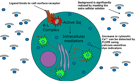
Calcium ion is an important element of the body. As a second messenger in the cell, it activates various protein kinases (such as adenylate cyclase, phosphodiesterase, calmodulin kinase, etc.) and many proteolytic enzymes by binding to calmodulin. And nucleases, which participate in the regulation of various cellular functions including cell metabolism, cell cycle, etc., play an important role in many life activities of cells. The traditional Fura-2, indo-1, and Quin-2 are Ca2+ fluorescent indicators that reflect changes in intracellular calcium concentration. When combined with calcium ions, the maximum excitation wavelength changes, the intensity of the emitted fluorescence and the bound Ca2+. The concentration has a quantitative relationship, but these calcium flow detection reagents have their own shortcomings. First, the entire reaction process requires water washing, which is not suitable for high-throughput experiments, and the second is sensitive to temperature and poor light stability. Molecular Devices has introduced a series of detection kits for calcium flow detection. Firstly, it uses the patented quench technology. The whole reaction process does not require water washing, increasing the detection flux. Secondly, it uses a special fluorescent probe to stabilize it after binding with calcium ions. The advantages of good performance and strong fluorescent signal have been affirmed by many experimenters.
Fluorescent protein reporter gene analysis
In recent years, in life science research, GFP (a green fluorescent protein extracted from jellyfish) has received more and more attention as a reporter gene with its own many advantages. First, GFP is non-toxic and does not require coenzyme itself. The light can be illuminated and the fluorescence signal is stable. We can use the gene of interest to fuse with the GFP gene, transfect the cells for expression, and determine the level of expression by the intensity of the fluorescent signal.
Time resolved fluorescence (TRF)
Time-resolved fluorescence (TRF) is actually developed on the basis of fluorescence intensity (FI), which is a special fluorescence analysis. Fluorescence analysis takes advantage of the large difference between the wavelength of the fluorescence and its excitation wavelength to overcome the influence of the variegated light in the ordinary ultraviolet-visible spectroscopic analysis. At the same time, the fluorescence analysis is different from the ordinary spectroscopic, the photoreceptor and the excitation light are not on the same line, and the excitation light The photoelectric receiver cannot be directly reached, which greatly improves the sensitivity of optical analysis. However, when ultra-micro analysis is performed, the influence of stray light of the excitation light becomes serious. Therefore, solving the influence of stray light of the excitation light becomes a bottleneck for improving sensitivity.
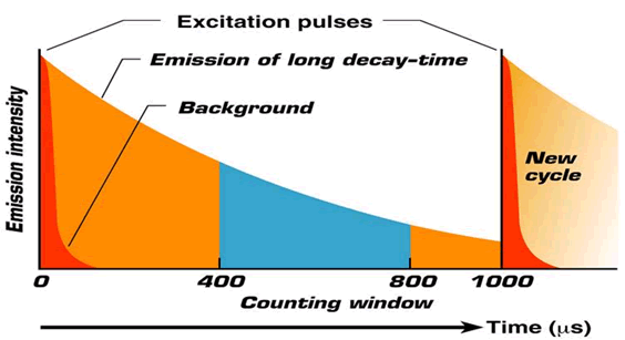
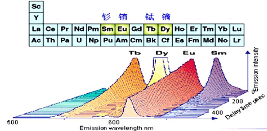
The best way to solve the effects of stray light is of course the absence of excitation light. However, the fluorescence lifetime of ordinary fluorescent markers is very short, the excitation light disappears, and the fluorescence disappears. However, the structure of the electron cloud of the trivalent ion of rare earth elements (Eu, Tb, Sm, Dy) limits the migration of electrons to some extent, and the decay period of the fluorescence of such elements is usually very long, and can reach 1- 2ms, can meet the measurement requirements, thus producing a time-resolved fluorescence analysis method, which is a method for analyzing the fluorescence intensity after the excitation light is turned off using a long-acting fluorescent label. The probes commonly used for time-resolved fluorescence detection are Eu (铕) and Tb (铽). Time-resolved fluorescence detection technology has the advantages of high sensitivity, strong specificity, good stability, good detection repeatability, short operation flow and wide standard curve range. It is suitable for micro-analysis of biological and medical immunology, such as cytokine detection. Antibody detection, viral antigen analysis, and drug metabolism analysis.
As people's requirements for food safety are getting higher and higher, the detection of toxic and harmful substances in food is becoming more and more strict in the international community, and the limit standards for various toxic and harmful substances are also increasing. Time-resolved fluorescence immunoassay methods are also available. For rapid detection of E. coli O157:H7, ochratoxin A, microcystins and harmful metals in foods. It is believed that with the wide application of multi-function microplate readers and the continuous development of various detection kits, time-resolved immunoassay technology will play a more powerful role in the field of food safety.
Fluorescence detection microplate reader
In summary, the use of fluorescent probes as detection means has many advantages, and more and more detection kits based on its principle are available. How can I better improve the sensitivity and linear range of the detection, reduce its background value, and improve the signal-to-noise ratio?
One of the key technologies to optimize the detection results, the core component of the microplate reader, the main function is to separate the narrow-band monochromatic light from a wide range of light. The earliest is the filter-type microplate reader, which is compatible with dichroic mirrors (in fact, another mode of filter reflective filter) and other optical path systems, which can meet the needs of most experiments. However, as the type of experiment changes and the detection requirements continue to increase, it is sometimes necessary to study the excitation and emission spectra. The filter-type microplate reader cannot meet the requirements due to the limitation of the number of filters, so the grating-type instrument was born. . The first grating-type microplate reader was introduced by Molecular Devices, which has a grating at each of the excitation and emission, but considering the insufficient purity of the light, a set of bands is added behind the excitation grating and the emission grating. The filter is blocked and the stray light is filtered twice, the result is basically the same as the pure filter system. After the patented technology of Molecular Devices is protected, many other microplate reader manufacturers can only use the double grating system on the grating type microplate reader to improve the light purity and reduce the stray light rate, that is, a set of gratings for excitation and emission. The whole optical path system has a total of four gratings, so it is also called a four-grating type microplate reader. Although it can also improve the stray light filtering, it has a large loss of light intensity due to the addition of a set of gratings, and the sensitivity of detection is also Will be affected.
The key technology to optimize the detection results, the choice of light source, the different transmittance of different monochromators to light, will affect the detection sensitivity, then the intensity of the light source itself will have a great impact on the experimental results, the current comparison of microplate readers Commonly used light sources are ordinary xenon flash lamps, high-energy xenon flash lamps and halogen lamps. LED light as a new light source is slowly adopted by many microplate readers with its own advantages. We know that under the same efficiency, LED lamp life is 10 times that of traditional halogen lamp, eliminating frequent replacement of bulb and waiting bulb. The trouble caused by preheating, the light source energy and stability are good, so it helps to improve the sensitivity of fluorescence detection.
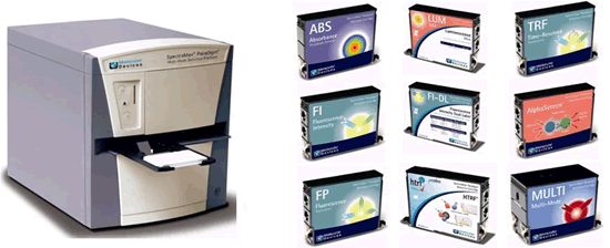
The traditional microplate reader adopts an integrated design mode of the optical path system and the detector system. The module is fixed at the factory and cannot be upgraded in function. There is a need for a multi-function microplate reader that users can upgrade at their own discretion. Molecular Devices has launched a multi-function microplate reader called Paradigm for market demand. It separates the light source system and the detection system independently. Multiple sets of detectors are fixed on the mainframe. Different cartridges are used for different experimental requirements. Each cartridge is actually an independent optical path system, such as optical absorption experiments in wavelength flexibility. For higher requirements, the light absorbing cartridge uses a grating as its monochromator; for the specificity of AlphaSreen requirements, the cartridge also uses a laser as its light source to ensure the reliability of the experimental data.
The only user on the market can upgrade the microplate reader
User upgrade time < 2 minutes
The launch of the Tune card box is considered to be another epoch-making breakthrough in the development of the microplate reader. It is a card box that combines the functions of fluorescence intensity, chemiluminescence and time-resolved fluorescence. First of all, it uses a high-energy AUTO-LED lamp as its light source, which not only increases the detection sensitivity, but also increases the linear range of its detection. The traditional microplate reader adjusts the fluorescent signal for different intensities in PMT (photomultiplier tube), fluorescence intensity. The linear range of detection is typically in the order of 4 to 5 orders of magnitude, and the AUTO-LED lamp can be used to increase the detection dynamic range to 6 to 7 orders of magnitude. Secondly, it combines the advantages of two different monochromators, using a new filter-adjustable technology, which is the flexibility of both the grating type monochromator (1nm light wave adjustable), and retains The filter-type monochromator features high light transmittance. Such fluorescence intensity and time-resolved fluorescence detection experiments not only increase the flexibility of detection but also increase the sensitivity of detection. Some data show that the sensitivity of the Tune cartridge is more than 10 times higher than that of the grating monochromator.
The sensitivity of the filter + the wavelength of the grating system is perfectly adjustable!

Molecular Devices has always focused on customer needs, and the introduction of the Paradigm multi-function microplate reader and Tune multi-function card box is a good example of this. For different requirements of different experiments in the future, Molecular Devices will launch a new target for it. The card box of the detection method is better to meet the needs of the user.
Anorectal Surgery Support,Sacrum Surgery Support,Operating Support Frame,Urology Surgery Accessories
NINGBO TECHART MEDICAL EQUIPMENT CO.,LTD , https://www.techartmed.com