For a given antigen, the optimal antibody concentration depends on the antigen and the antibody itself. The affinity of the antigen and the antibody, as well as the specific response of the primary antibody to the secondary antibody are different. Optimal antigen and antibody concentrations can be determined by immunoblotting experiments using different concentrations of antigen and antibody. In addition, a faster and simpler method is to perform a dot blot experiment.
The following is the method of operation of a dot blot using an ultrasensitive signal substrate:
1. A protein sample diluted with TBS and PBS. The good dilution method is determined by the dilution concentration of the antigen in the sample. However, since the concentration of the antigen is unknown, it is necessary to perform a wide range of dilution detection. The detection sensitivity of the SuperSignal West Pic chemiluminescent substrate is up to the skin g level, so the dilution range of the sample can range from microgram level to picogram level. If too many antigens are to be used, the result is the following: non-specific bands, blurred bands, and reduced signal.
2. Prepare the transfer film. The number of membranes required depends on how many different dilution concentrations are applied to the primary and/or secondary antibodies being shielded. Typically, the dilution of one or both primary antibodies is determined using two or three different secondary antibody dilutions. For example: 1/1000 primary antibody and 1/50000 secondary antibody; 1/1000 primary antibody and 1/100000 secondary antibody; 1/5000 primary antibody and 1/50000 secondary antibody; and 1/5000 and 1 1.000000 secondary antibody.
3. Place the film on the filter paper. The antigen dilution was spotted on the membrane. Use as small a dilution as possible on the membrane (2-5 ul is preferred), as the larger the volume, the more diffuse the signal. The antigen solution is dried on the membrane for 10-30 minutes or until no visible moisture is present.
4. Block the non-specific spots on the membrane with 0.05% Tween-20 blocking solution and incubate for 1 h at room temperature.
5. Dilute the primary antibody into the blocking solution/Tween-20 detergent and add to the membrane and incubate for 1 h at room temperature.
6. Wash the membrane 4-6 times with TBS or PBS, and add 0.05% Tween to the eluate with the largest possible volume of eluent to reduce the non-specific background. During each elution, the membrane was suspended in the eluate for about 5 minutes, and the eluate was poured out and repeated. A short rinse in the eluent may increase the elution efficiency before incubation.
7. Prepare a secondary antibody/HRP-conjugated blocking reagent/Tween-20 detergent diluent, and add the secondary antibody dilution to the membrane for 1 h.
8. Clean the membrane again by following the procedure in step 6.
9. Prepare substrate working buffer, mix equal volumes of luminol/enhancer and stabilized peroxide solution. Prepare enough volume to ensure that the blot is completely wet, ensuring that the blot does not dry during the incubation. Recommended volume: 0.1 ml/cm2 blotting surface.
10. Incubate the membrane in a super-sensitive chromogenic substrate working solution for 5 minutes;
11. Remove the film from the substrate and place it on a plastic sheet or other protective film;
12. Attach the blot to the film and expose it next to the protein. Any standard or enhanced autoradiographic film can be used. The first exposure is recommended for 30-60 s, and the exposure time can be different for best results. In addition, CCD cameras or other imaging facilities may be used; however, these facilities may require longer exposure times.
13. On the best blotting membrane, the signal generated by the super-sensing reaction substrate may last for more than 8 h. Without the best results, the blot can be repeatedly exposed to film or to an imaging system. As the blotting time increases, the exposure time needs to be extended. If the best results are not obtained, this procedure can be repeated with antigen and/or antibody dilutions.
Related reagents for this experiment:
1, chemical reagents: TBS, PBS, Tween-20, luminol, hydrogen peroxide solution, etc.
2. Biological reagents:
Corresponding primary antibody applied to immunospot assay
HRP enzyme secondary antibody such as rabbit source, mouse source, sheep source, and Wuyuan
Rabbit enzyme, mouse source, sheep source, Wuyuan and other AP enzyme standard secondary antibodies
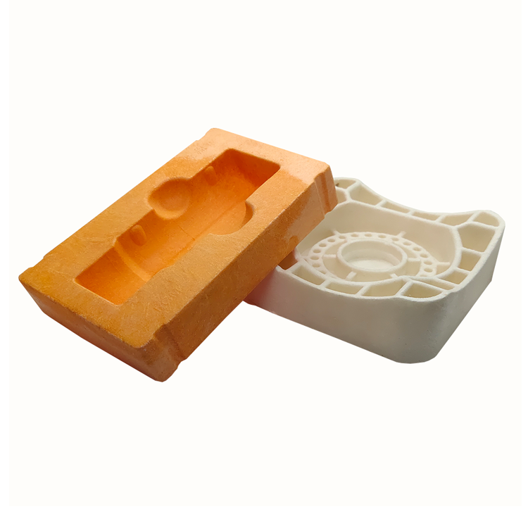
Biodegradable Materials Blister Tray
Degradable Blister Box,Biodegradable Blister Packaging,Biodegradable Blister Plastic Tray,Biodegradable Materials Blister Tray
1, with excellent transparency and smoothness, display effect is good.
2, the surface decoration performance is excellent, can be printed without surface treatment, easy to press the pattern, easy to metal treatment (vacuum gold-plated layer)
3. It has good mechanical strength.
4. Good barrier performance for oxygen and water vapor.
5, good chemical resistance, can withstand a variety of chemical substances corrosion.
6, non-toxic, reliable health performance
7, good adaptability to environmental protection, can be economical and convenient recycling; When the waste is incinerated, it does not produce harmful substances harmful to the environment.

Degradable Blister Box,Biodegradable Blister Packaging,Biodegradable Blister Plastic Tray,Biodegradable Materials Blister Tray
taicang hexiang packaging material co.,ltd , https://www.medpackhexiang.com

