Release date: 2009-11-20
Four-dimensional ultrasound has a variety of imaging modes, which has a certain significance in the diagnosis of fetal congenital heart disease. It can show the spatial shape, orientation and relationship with the surrounding tissues, making it easier for doctors and pregnant women to understand. It also simplifies the process of image acquisition. Reduced reliance on operational experience.
1. Multi-planar method (multi2planar, MP) is to display the morphological structure of the observed part from three mutually perpendicular X, Y, Z two-dimensional sections, which are called A, B, C planes respectively, where A plane and probe The directions are parallel, the B plane is parallel to the sound beam and perpendicular to the A plane, and the C plane is perpendicular to the sound beam. The examiner can adjust the volume database by moving the X, Y, Z axes and orthogonal points of the three planes to display any of the sections in the volume library, and to display the morphological structure of the heart in different directions.
2. Tomograp hic ult rasound im2aging (TUI) is a multi-planar analysis method similar to magnetic resonance or CT imaging. It can simultaneously display 9 consecutive parallel slices in the volume database. These slices are from - 4 Arrange to + 4, where 0 is the original acquisition slice and the top left is the sagittal map. The examiner can adjust most of the sections required for fetal echocardiography by adjusting the layer spacing of the section. The researchers used TUI to retrospectively analyze 103 fetal hearts diagnosed as congenital heart disease. By adjusting the layer spacing, the vascular morphology and connection of the large vessels can be displayed on different sections, and segmental analysis is performed. Check the difficulty of congenital heart disease.
3. Surface rendering is ideal for fetal facial imaging and has also been used in four-dimensional ultrasound imaging of fetal heart in recent years. The examiner places the sampling frame in the volume of interest to demonstrate the dynamic three-dimensional structure of the volume, such as the interatrial septum, interventricular septum, trabecular bone, foramen ovale, and atrioventricular valve. Using this technique, the researchers compared 38 cases of fetal heart malformation with 136 normal fetal hearts. The results showed that the normal fetal heart interval and ventricular septal rate were 9613 %, and the atrioventricular annulus was 9314%. In the fetal heart In the deformed group, the morphological structure and motion state of the foramen ovale were observed in 4 cases; atrioventricular annulus imaging was performed on the coronal section, and 5 cases showed the relationship between the beginning of the aorta and the atrioventricular valve, and 3 cases could be used. To evaluate the semilunar valve and observe the presence or absence of macrovascular abnormalities. The imaging method provides a completely different perspective from the two-dimensional imaging to observe the structure of the atrial, interventricular septum and annulus, and is conducive to the diagnosis and understanding of fetal congenital heart disease.
4. Glass body mode Four-dimensional ultrasound can be combined with color or energy Doppler technology to obtain a volume database with blood flow signals when collecting volume. By adjusting the threshold of the grayscale and color scale of the image and the setting of the transparency, the grayscale of the blood flow surface is translucent, showing the blood flow inside. The examiner can generally observe and evaluate the vascular structure of the great vessels of the heart, and can reconstruct the vascular tree in the chest and abdomen of the fetus. It is not necessary to collect a large number of two-dimensional sections to confirm the type of vascular malformation.
5. Invert sion imagine mode is a post-processing mode that can be combined with STIC technology to analyze the volume database. The mode displays the echo generated by the tissue as colorless, and the liquid is displayed as strong echo. Myocardium is shown as no echo, while blood flow is shown as white. In addition, it has the advantage of rebuilding the stomach bubble and gallbladder, which helps to diagnose the complex malformation of the heart.
6. After the volume data is acquired by the four-dimensional ultrasound, it can be stored in the hard disk of the machine, and then used for offline analysis or teaching use; the volume database can be shared through the network to promote remote consultation and interdisciplinary communication. Shanghai Medical Device Industry Association
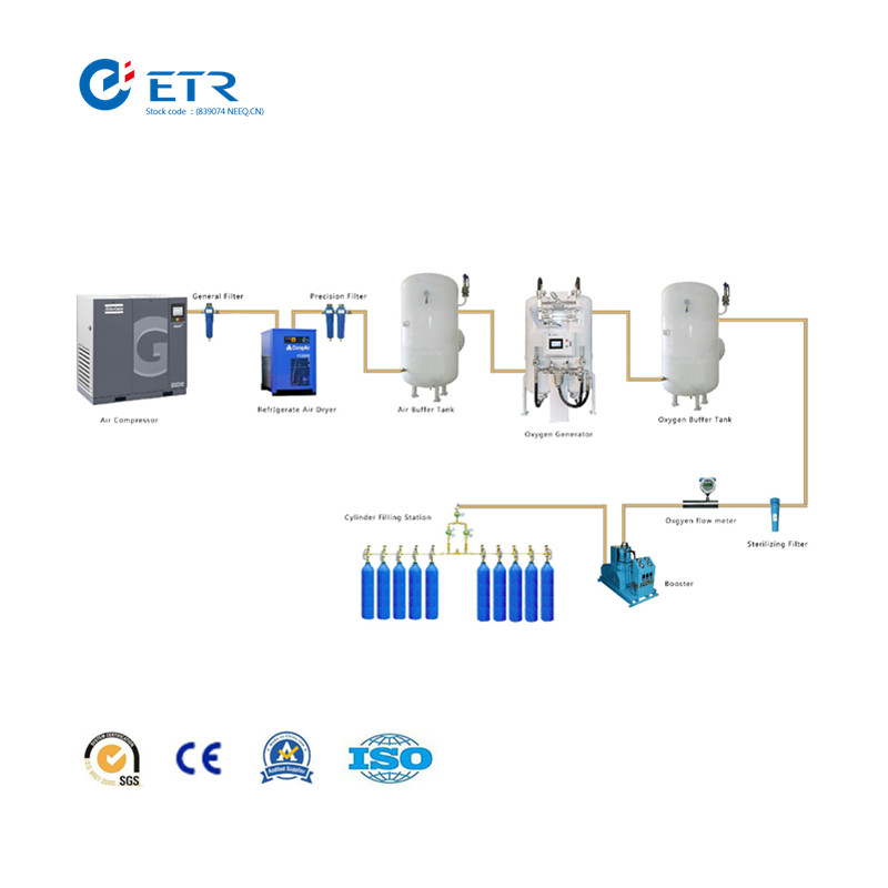
ETR Oxygen Cylinder Filling System
By PSA principles, ETR PSA oxygen generators generate 93%±2% purity oxygen gas directly from compressed air. ETR oxygen cylinder filling plant is consisted of Atlas Copco air compressor, Parker dryer and filters, air buffer tank, ETR Psa Oxygen Generator, oxygen buffer tank, oxygen receiver tank, diaphragm oxygen booster, filling station and control cabinet.
Compressed air is purified through the air dryer and filters to a certain level for main generator to work with. Air buffer is incorporated for smooth supply of compressed air thus to reduce fluctuation of compressed air source. The generator produces oxygen with PSA (pressure swing adsorption) technology, which is a time proven oxygen generation method. Oxygen of desired purity at 93%±2% is delivered to oxygen buffer tank for smooth supply of product gas. Oxygen in buffer tank is maintained at 4bar pressure, then reach to 150bar to filling cylinders with an oxygen booster.

Oxygen Cylinder Filling System
Oxygen Cylinder Filling System,Homefill Oxygen System,Oxygen Refilling Plant,Oxygen Refilling Station
Hunan Eter Electronic Medical Project Stock Co., Ltd. , https://www.centralgas.be
